CE EMG Needle Electrode Length: 28mm - Durable and Reliable
Product Name :EMG Needle Electrode
Registered Trademark: Lingan Brand
Product use and scope of application: This product is mainly used in the field of neurophysiology, by neurologists to record the human muscle-related electrical activity, and is recommended to be used in conjunction with electromyography machines.
| Style |
Length |
Needle diameter |
Recorded area |
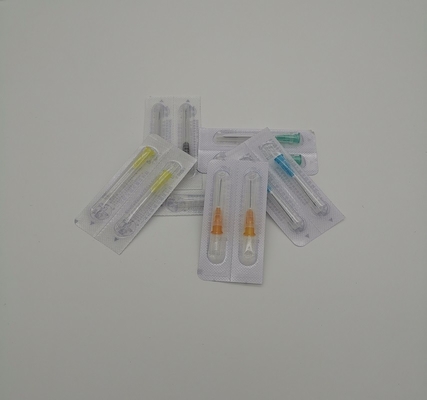
Color |
Packaging |
| CNE35-25 |
25mm |
0.35mm |
0.03mm² |
Pink |
25pcs/Box |
| CNE45-28 |
28mm |
0.45mm |
0.07mm² |
Red |
25pcs/Box |
| CNE45-38 |
38mm |
0.45mm |
0.07mm² |
Orange |
25pcs/Box |
| CNE45-50 |
50mm |
0.45mm |
0.07mm² |
Black |
25pcs/Box |
| CNE50-60 |
60mm |
0.50mm |
0.07mm² |
Yellow |
25pcs/Box |
Electromyography is an electrophysiological examination in which needle electrodes are inserted into the muscle to record changes in potential.
The signal of skeletal muscle contraction is the action potential (AP) of the cell, which can be conducted by volume conductors on the cell and recorded by extracellular electrodes or body surface electrodes
EMG in a narrow sense refers exclusively to concentric needle EMG/conventional EMG.
The identification of EMG needs to be combined with waveform and sound, and this article analyzes common EMG potentials with respect to waveform.
The clinical application of conventional EMG is to diagnose and differentiate neurogenic damage from myogenic damage.
EMG records three main phases of EMG signals: resting, light contraction and heavy contraction.

Four steps of needle polar electromyography observation
①Insertion of electrical activity: The change in potential caused when the recording needle is inserted into the muscle.
②When relaxed: observe whether there is abnormal spontaneous electrical activity in the muscle when it is completely relaxed.
③When contracting lightly: observe the motor unit potential time frame, wave amplitude, phase and frequency of issue.
④ During vigorous contraction: Observe the type of motor unit potential raising.
Since EMG activity (in microvolts) is linearly correlated with the amount of muscle contraction and the number of contracting muscles, in other words, the more intense the muscle contraction, the greater the number of motor units in the activated muscle, and the higher the recorded voltage amplitude.
What is electromyography?
1. Using a myoelectric needle to pierce a muscle and observe the bioelectric changes in the muscle in different states.
2. Using pulsed current, stimulate the nerves in different parts of the body and observe the bioelectric changes in the nerves and their innervated muscles.
3. To reflect the functional state of the nerve muscles

 Your message must be between 20-3,000 characters!
 English