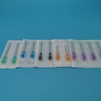
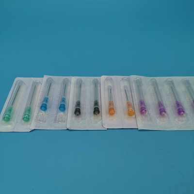
CE Certified Ergonomic Hub EMG Needle Electrode Length: 38mm
What is electromyography?
1. Using a myoelectric needle to pierce a muscle and observe the bioelectric changes in the muscle in different states.
2. Using pulsed current, stimulate the nerves in different parts of the body and observe the bioelectric changes in the nerves and their innervated muscles.
3. To reflect the functional state of the nerve muscles.
The needle electrode used in EMG examination is called disposable needle electrode (aka: myoelectric needle, concentric needle), which is a bipolar fine needle electrode. The electrode is inserted into the muscle during the examination, and the bio-current of the muscle in the resting and contracted states is amplified by a signal amplifier and then displayed by an oscilloscope.
Features】: 1:
1. Needle: smooth needle cut surface, smooth needle entry, patient pain is small.
2. Needle: good flexibility, laser-cut surface, smooth and burr-free.
3. Connector: gold-plated at the connector to reduce signal distortion, signal stability, high accuracy.
4. Handle: non-slip design, plastic projection marking the direction of the needle tip cross-section, easy to use.
Product use and scope of application: This product is mainly used in the field of neurophysiology, by neurologists to record the human muscle-related electrical activity, and is recommended to be used in conjunction with electromyography machines.
| Style |
Length |
Needle diameter |
Recorded area |
Color |
Packaging |
| CNE35-25 |
25mm |
0.35mm |
0.03mm² |
Pink |
25pcs/Box |
| CNE45-28 |
28mm |
0.45mm |
0.07mm² |
Red |
25pcs/Box |
| CNE45-38 |
38mm |
0.45mm |
0.07mm² |
Orange |
25pcs/Box |
| CNE45-50 |
50mm |
0.45mm |
0.07mm² |
Black |
25pcs/Box |
| CNE50-60 |
60mm |
0.50mm |
0.07mm² |
Yellow |
25pcs/Box |

Electromyography is an electrophysiological examination in which needle electrodes are inserted into the muscle to record changes in potential.
The signal of skeletal muscle contraction is the action potential (AP) of the cell, which can be conducted by volume conductors on the cell and recorded by extracellular electrodes or body surface electrodes
1. Resting state
The following two types of potentials can be observed in the resting state:
(1) Insertion potential: a brief potential release when the needle electrode is inserted into the muscle, which disappears when the needle feeding is stopped.
Prolonged or increased insertion potentials are seen in neurogenic and myogenic damage; decreased or absent insertion potentials are seen in muscle fibrosis and adiposity.
(2) Spontaneous potentials: fibrillation potentials, positive sharp waves, fasculations, myokymic discarges and complex repetitive discarges (CRD), etc.


 Your message must be between 20-3,000 characters!
 Please check your E-mail!
  Your message must be between 20-3,000 characters!
 Please check your E-mail!


